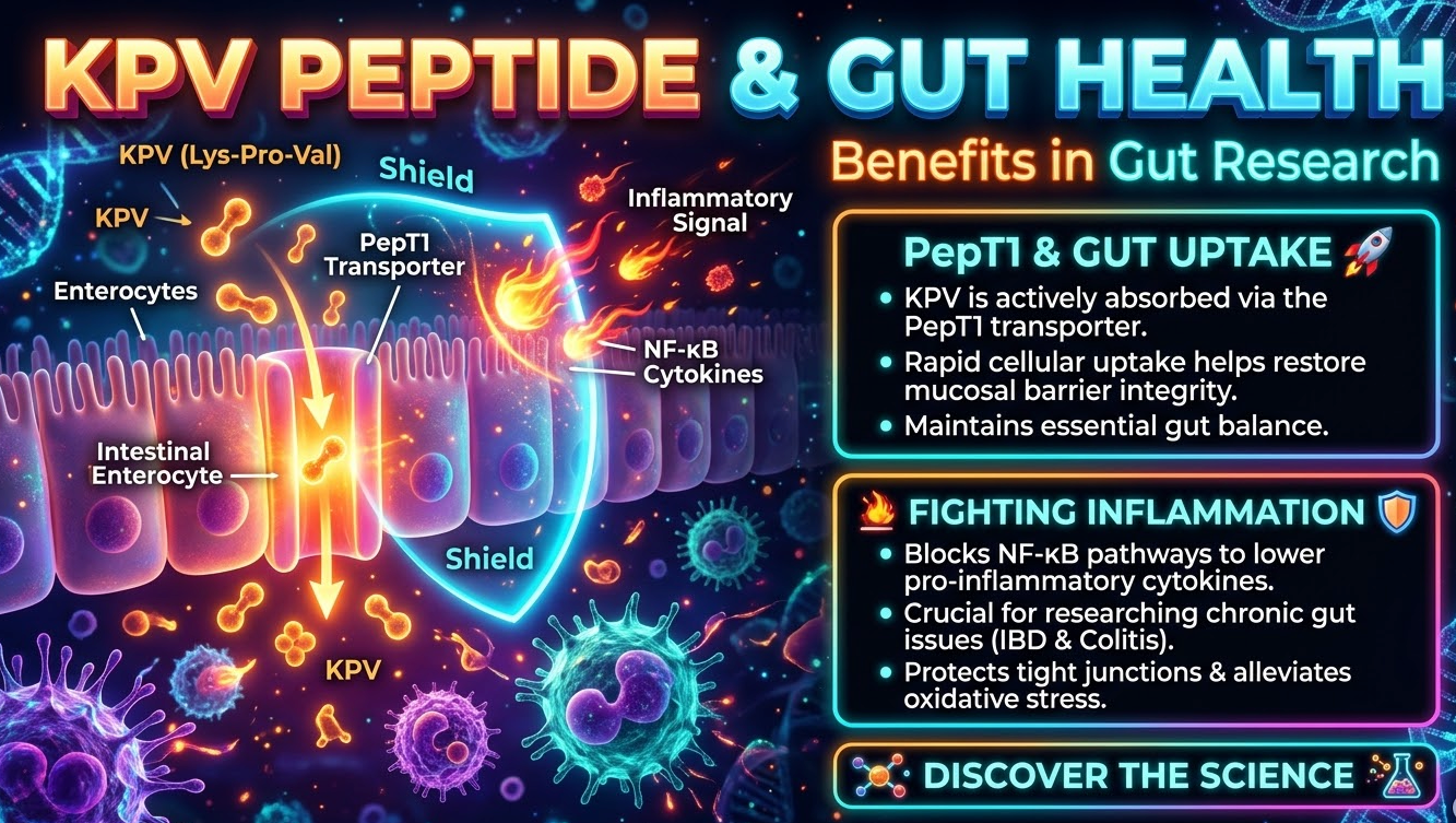
Delivering intact biological molecules past the digestive system's enzymatic barriers is one of the most formidable hurdles in gastrointestinal medicine. While larger peptides degrade into inactive amino acids when swallowed, KPV (Lysine-Proline-Valine) bypasses digestive breakdown by utilizing a specialized active cellular gateway: the PepT1 intestinal transporter. Understanding KPV PepT1 gut research explains how this tiny tripeptide enters colonocytes directly to suppress NF-κB inflammatory cascades, restore mucosal integrity, and modulate inflammatory bowel models.
⚡ Quick Summary: KPV PepT1 Gut Research & Mechanisms
Quick Answer: KPV is an ultra-small, three-amino-acid C-terminal fragment of alpha-melanocyte-stimulating hormone (α-MSH). Unlike larger regenerative peptides that break down in digestive enzymes, KPV is actively imported into intestinal epithelial cells via the PepT1 (Peptide Transporter 1) carrier. Inside the colonocyte, KPV translocates into the nucleus to block NF-κB activation, drastically suppressing pro-inflammatory cytokines (TNF-α, IL-1β, and IL-6) and accelerating mucosal healing in IBD and colitis research models.
| Research Parameter | KPV Biological Profile | Laboratory Relevance |
|---|---|---|
| Molecular Size & Weight | Tripeptide (Lys-Pro-Val), ~383.5 Da | Under 500 Da threshold; evades enzymatic proteolysis |
| Cellular Influx Pathway | Active PepT1 symporter (H+-coupled) | Overexpressed in inflamed colonic mucosa for targeted uptake |
| Intracellular Target | Nuclear Factor Kappa B (NF-κB) | Prevents p65 translocation, arresting cytokine cascades |
| Delivery Modalities | Oral (entero-targeted), SubQ, or Topical | Oral delivery directly targets gut lumen; SubQ targets systemic tissue |
The PepT1 Gateway: How KPV Reaches Intestinal Epithelium
In a healthy gut, PepT1 (SLC15A1) is an apical proton-coupled di- and tripeptide transporter predominantly active in the small intestine. However, in pathological states like Crohn’s disease, ulcerative colitis, and chronic gut dysbiosis, PepT1 is drastically upregulated in colonic epithelial cells and immune cells.
1. Active Transporter Influx
Because KPV is exactly three amino acids long, it fits the precise substrate criteria for PepT1, enabling rapid cellular entry without requiring destructive pinocytosis.
2. Targeted Pathological Affinity
Because inflamed colonocytes overexpress PepT1, KPV naturally concentrates at the precise tissue sites experiencing the highest inflammatory stress.
3. Direct NF-κB Inactivation
Once inside, KPV suppresses the phosphorylation and degradation of IκB-α, trapping NF-κB outside the nucleus and stopping the transcription of destructive inflammatory mediators.
Advance Your Gut Barrier & Inflammation Research
Investigate the potent anti-inflammatory mechanisms of pure Lys-Pro-Val. Discover certified analytical purity and laboratory-verified specifications for KPV.
Shop Research KPV →Delivery Dynamics: Oral vs. Subcutaneous vs. Nanoparticle Carriers
A critical consideration in KPV PepT1 gut research is distinguishing local luminal exposure from systemic distribution. Route selection dictates the experimental outcome:
- Oral & Enteric Targeting: Oral administration places KPV in direct contact with the apical membrane of intestinal cells where PepT1 is active. In published animal colitis studies, oral administration demonstrated superior localized mucosal repair compared to systemic routes (Dalmasso et al., 2008).
- Nanoparticle Encapsulation: Cutting-edge research utilizes biocompatible nanoparticles (such as hyaluronic acid or PLGA carriers) to encapsulate KPV, shielding it from gastric acid and releasing it specifically in the inflamed colon (Laroui et al., 2010).
- Subcutaneous Administration: SubQ injection bypasses the gut lumen, distributing KPV into systemic circulation to target joint inflammation, dermatitis, and systemic immune overactivation.
Synergistic Research Pairings for Gut & Tissue Repair
Modern regenerative protocols often combine KPV with complementary tissue-healing compounds to evaluate multi-pathway gut barrier restoration:
- The Multi-Target Formulation: Explore KLOW Blend, which unites KPV with BPC-157, TB-500, and GHK-Cu for comprehensive mucosal, vascular, and tissue matrix repair. Read our breakdown: GLOW vs. KLOW Peptide: Comparing the 70mg & 80mg Blends.
- The Healing Gold Standard: Compare localized KPV cellular uptake with systemic angiogenic repair in our Wolverine Blend (BPC-157 / TB-500). Review the dosing mechanisms: What is the Wolverine Blend? Research & Dosing Protocols.
- Gut Axis & Metabolic Dysbiosis: Severe gut inflammation often coexists with metabolic dysregulation. Learn how gut hormones influence mucosal pathways in GLP-1 and the Gut: Navigating IBS, Crohn's & Colitis.
Standardized 2.0 mL Reconstitution Math for Research Vials
When preparing injectable KPV or setting up benchtop assay stock, accurate diluent measurement is mandatory. Below is the standardized 2.0 mL reconstitution calculation using certified Pfizer Hospira Bacteriostatic Water:
Concentration Formula: Mass (mg) ÷ Diluent Volume (mL) = Concentration (mg/mL). For example, dissolving 10 mg of lyophilized KPV in 2.0 mL of bacteriostatic water yields an active concentration of 5 mg/mL (500 mcg per 0.1 mL / 10 units on a U-100 syringe).
| Vial Material Amount | Diluent Added (BAC Water) | Final Concentration | Dose per 10 Units (0.1 mL) |
|---|---|---|---|
| 10 mg Vial | 2.0 mL | 5.0 mg/mL (5,000 mcg/mL) | 500 mcg (0.50 mg) |
| 20 mg Vial | 2.0 mL | 10.0 mg/mL (10,000 mcg/mL) | 1,000 mcg (1.00 mg) |
| 50 mg Vial | 2.0 mL | 25.0 mg/mL (25,000 mcg/mL) | 2,500 mcg (2.50 mg) |
Calculate custom volume conversions and unit fractions instantly with the free interactive Peptide Calculator.
Hardware Selection: Moving Beyond Disposable Syringes
Maintaining high experimental precision requires hardware that shields reconstituted peptides from environmental shock and delivers exact volume fractions:
- Reusable Injection Pens: For systemic injectable protocols, transfer your dissolved KPV solution into 3mL Injection Pen Glass Cartridges and pair with the V3 Reusable Injection Pen. The mechanical dial eliminates syringe parallax errors and prevents compound loss in needle hubs.
- Precision Manual Syringes: If manual draws are required, select ultra-fine Easy Touch 31G U-100 Syringes with 0.5 mL barrels, which offer wide graduation spacing for micro-volume precision without coring rubber stoppers.
- Secure Lab Storage: Keep your reconstituted KPV vials indexed, upright, and light-shielded inside the Compact 4-Slot Case or the high-capacity 10-Slot Peptide Vial Case. For mobile protocols, secure supplies within the insulated Compact Peptide Travel Case.
Protocol Builder & Tracking Tools
Document every step of your KPV research to ensure consistent reproducibility:
- Protocol Structuring: Plan your multi-week compounding timelines and delivery schedules using the Protocol Builder.
- Observation Logging: Track barrier integrity indicators, beyond-use dates, and subject metrics using the Protocol Tracker Tool.
Frequently Asked Questions
Is oral KPV research automatically applicable to subcutaneous injections?
No. While subcutaneous KPV produces potent systemic anti-inflammatory effects, oral delivery specifically leverages apical PepT1 transporters expressed on inflamed gut epithelial cells. The route of delivery fundamentally alters whether the peptide acts primarily within the intestinal lumen or throughout systemic vascular beds.
Does KPV carry the same pigment-darkening effects as parent α-MSH?
No. Full-length α-MSH binds to melanocortin 1 receptors (MC1R) to trigger skin pigmentation. KPV represents only the C-terminal tripeptide fragment; it retains the potent anti-inflammatory and NF-κB inhibitory actions of α-MSH without activating melanogenesis or causing skin darkening.
How should reconstituted KPV be stored?
Lyophilized KPV cakes are stable at room temperature for short periods, but once reconstituted with bacteriostatic water, the solution must be stored refrigerated between 2°C and 8°C. Keep the vial protected from direct light and mechanical shaking to preserve tripeptide stability.
Related Reading & Scientific References
- KPV Peptide Explained: Gut Barrier Action, Dosing Protocols & Research
- GLP-1 and the Gut: Navigating IBS, Crohn's, and Colitis
- Dalmasso et al. (2008): PepT1-Mediated Tripeptide KPV Transport in Intestinal Inflammation (PubMed)
- Laroui et al. (2010): Colon-Targeted Nanoparticles for KPV Delivery in Murine Colitis (PubMed)
The ability of KPV to enter inflamed colonocytes via PepT1 transporters makes it a cornerstone molecule in modern gastrointestinal research. By targeting NF-κB signaling at the nuclear level, KPV opens promising experimental avenues for mucosal restoration and inflammatory bowel resolution.
← View all peptide guides, research & protocols in our Blog Archive