KPV Peptide Explained: Gut Barrier Action, Dosing Protocols & Research
In gastrointestinal research and dermatological science, investigators view KPV as a targeted mucosal protector. This short tripeptide calms aggressive immune signaling without causing the broad immunosuppression typical of conventional pharmaceuticals.
Online wellness forums often describe KPV as an instant fix for irritable bowel disorders, systemic histamine reactions, and mast cell activation. However, researchers must separate marketing enthusiasm from verified biochemistry. Understanding KPV requires examining its origins as an alpha-MSH fragment, its active transport mechanisms, and its performance across controlled mucosal barrier trials.
Quick Summary: What Is KPV at a Glance?
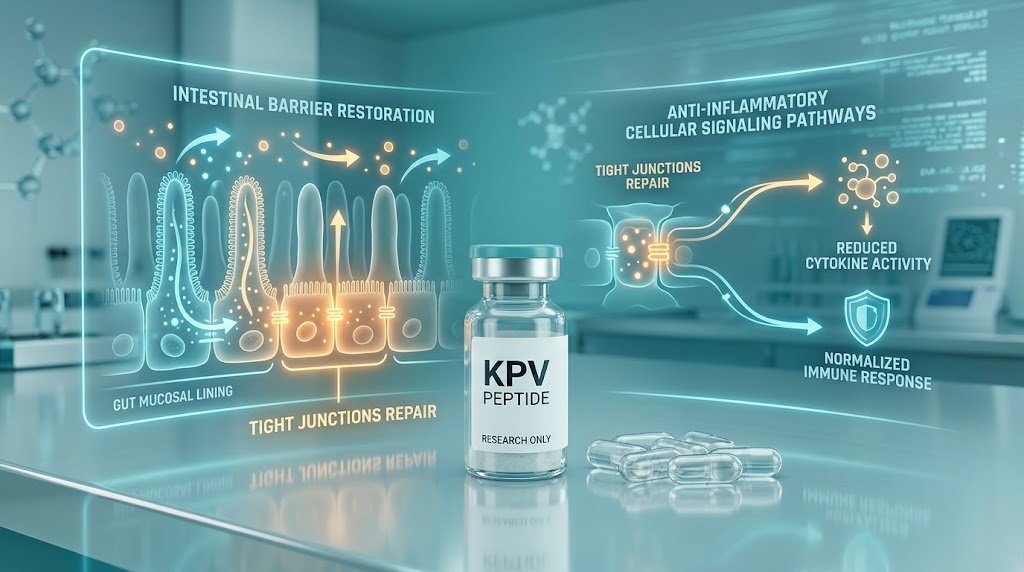
Quick Answer: KPV is a synthetic tripeptide (Lysine-Proline-Valine) representing the C-terminal sequence of Alpha-Melanocyte-Stimulating Hormone (α-MSH). It suppresses mucosal inflammation by utilizing PepT1 transporters to enter epithelial cells, migrating into the nucleus to inhibit NF-κB activation, stabilizing epithelial tight junctions, and exhibiting direct antimicrobial properties against pathogens like Candida albicans—all without stimulating pigment-producing MC1R receptors.
| Parameter | Oral Research Protocol | Subcutaneous (Sub-Q) Research |
|---|---|---|
| Primary Focus | Direct gastrointestinal mucosal contact, IBD/colitis models, gut lining defense | Systemic inflammation, autoimmune modulation, dermatological and joint calming |
| Typical Evaluation Dose | 200–500 mcg daily (taken in a fasted state) | 200–500 mcg daily |
| Primary Mechanism | PepT1 active transport into mucosal epithelial cells, tight junction stabilization | Direct nuclear translocation, NF-κB promoter blockade, systemic cytokine suppression |
| Direct Research Supply | Lyophilized high-purity research vials available via KPV (5mg / 10mg) | |
What Is KPV?
KPV is a naturally derived tripeptide containing three amino acids: Lysine-Proline-Valine.
Its sequence matches the exact C-terminal end of Alpha-Melanocyte-Stimulating Hormone (α-MSH), a 13-amino-acid peptide that the pituitary gland and peripheral immune cells generate naturally.
While full-length α-MSH reduces inflammation effectively, it also binds strongly to melanocortin-1 receptors (MC1R), which triggers melanin production and darkens skin tissue. Biochemists synthesized the isolated KPV fragment specifically to preserve the parent molecule's potent anti-inflammatory and antimicrobial actions while bypassing the pigment-producing pathways entirely.
Mechanisms of Action: How KPV Works at the Cellular Level
KPV avoids conventional surface receptors. Instead, it enters target cells directly to modify core inflammatory transcription pathways:
1. Active Influx via the PepT1 Transporter
Most large peptides cannot easily cross cellular membranes. KPV bypasses this hurdle by utilizing PepT1 (Peptide Transporter 1), an active transport protein lining intestinal epithelial cells. Inflamed mucosal tissues upregulate PepT1 heavily, allowing KPV to target damaged epithelial sites rapidly and concentrate where tissues face the greatest stress.
2. Direct Nuclear Inhibition of NF-κB
Once inside the cytoplasm, KPV moves straight into the nucleus. It prevents Nuclear Factor kappa B (NF-κB) from translocating and binding to genomic promoter regions. By halting this key genetic switch, KPV suppresses the production of destructive pro-inflammatory cytokines, including TNF-α, IL-1β, and IL-6.
3. Epithelial Tight Junction Stabilization
During intestinal inflammation, damaged barrier walls let pathogens pass into deeper tissue layers. In mucosal injury models, KPV preserves critical tight-junction proteins like occludin and claudins. This action seals the intercellular gaps, limits inflammatory cell infiltration, and maintains epithelial structure.
4. Direct Antimicrobial Membrane Lysis
Beyond regulating cytokines, KPV exhibits direct antimicrobial actions against common opportunistic pathogens. In vitro assays demonstrate that KPV disrupts cell walls and halts colony growth in microbes like Staphylococcus aureus and Candida albicans, protecting host tissues from microbial overgrowth.
What Does the Research Actually Show?
A rigorous assessment of KPV requires weighing encouraging cellular and animal data against the scarcity of controlled human trials.
Compelling Animal Colitis Literature
Published KPV literature centers primarily on rodent models of inflammatory bowel disease, including DSS- and TNBS-induced colitis. Across these animal studies, both oral and injectable KPV reliably reduced mucosal ulceration, preserved colon length, and prevented rapid body weight loss.
The Clinical Data Gap
Despite impressive animal findings, researchers have yet to conduct large-scale, double-blind, placebo-controlled clinical trials using pure synthetic KPV in human patients. Most public reports rely on functional medicine case notes and observational wellness protocols rather than formal Phase III clinical datasets.
Regulatory Standing
Health regulatory bodies, including Health Canada and the US FDA, have not approved KPV to treat, diagnose, or prevent human diseases. It remains an investigational research peptide designated exclusively for laboratory experimentation.
Documented Laboratory Research & Dosing Protocols
Published literature, animal trials, and laboratory models document the following experimental parameters. When planning syringe volume tick marks, utilize our free Peptide Calculator and track multi-week administration intervals via the Protocol Tracker Tool.
| Parameter | Subcutaneous (Sub-Q) Protocol | Topical / Dermal Protocol |
|---|---|---|
| Experimental Focus | Systemic inflammation, autoimmune gut models, mucosal recovery | Dermal inflammation, wound calming, localized skin irritation |
| Documented Dosing | 200–500 mcg per day | 0.3%–0.5% concentration, applied 1–2 times daily |
| Administration Route | Subcutaneous (Sub-Q) injection | Applied directly onto the target dermal area |
| Cycle Duration | 4–8 weeks continuous evaluation (review our Peptide Cycle Timing Guide) | 4–8 weeks continuous application |
| Washout Window | 2–4 weeks off between experimental cycles | 2–4 weeks off between cycles |
| Screening Panels | CMP, C-Reactive Protein (CRP), Complete Blood Count (CBC) | Baseline patch testing, localized skin evaluation, CMP |
Oral vs. Subcutaneous Administration in Research
Because PepT1 transporters line the brush border of the small intestine and colon, researchers frequently select oral liquid or capsule routes when investigating direct gastrointestinal contact. Conversely, studies targeting systemic autoimmune signaling or non-gut tissues prefer subcutaneous injections to ensure reliable systemic bioavailability.
Synergistic Laboratory Pairings & Multi-Peptide Blends
Researchers often evaluate KPV alongside complementary peptides to study multi-target mucosal and connective tissue defense:
- BPC-157: Scientists frequently pair KPV with BPC-157 to analyze combined tight-junction preservation and VEGFR2-mediated blood vessel repair.
- TB-500: Researchers combine KPV with TB-500 to evaluate actin-driven cell migration alongside direct NF-κB inhibition.
- Wolverine Blend: Teams compare KPV against the dual-pathway tissue remodeling action of the Wolverine Blend Guide (or review calibrated single-solution vials on the Wolverine Blend product page).
- KLOW Blend (80mg): Unites KPV (10mg) with GHK-Cu (50mg), BPC-157 (10mg), and TB-500 (10mg) in a single solution for advanced mucosal and deep tissue repair. Explore the KLOW Blend or read our GLOW vs. KLOW Comparison Guide.
- GHK-Cu: Dermal research teams study KPV with GHK-Cu Copper Peptide (available as standalone GHK-Cu 50mg/100mg) to evaluate extracellular matrix remodeling alongside localized anti-inflammatory calming.
Laboratory Handling, Reconstitution & Storage Standards
Its short three-amino-acid structure makes KPV resilient, yet maintaining experimental precision requires standard laboratory care (detailed step-by-step in our Beginner's Guide to Peptides):
- Diluent Selection: Reconstitute lyophilized KPV using sterile, pharmaceutical-grade Pfizer Hospira Bacteriostatic Water containing 0.9% benzyl alcohol to prevent bacterial growth during multi-week protocols. Alternatively, use 0.9% Sterile Bacteriostatic Saline to maintain isotonic conditions.
- Reconstitution Protocol: Use a sterile EasyTouch 31G Syringe to direct diluent slowly down the vial's interior glass wall. Do not spray diluent directly onto the lyophilized cake. Roll the vial gently between your palms until the powder dissolves; avoid violent shaking. Standard 10 mg research vials accommodate up to 3 mL of diluent.
- Cold-Chain Storage: Store unmixed lyophilized vials at -20°C for long-term stability. Maintain reconstituted liquid solutions between 2°C and 8°C (36°F to 46°F) inside a secure, light-shielded Peptide Vial Case or insulated Compact Travel Case to protect against light exposure and mechanical agitation.
KPV provides an effective, targeted model for anti-inflammatory peptide research. By delivering the functional C-terminal sequence of α-MSH directly into cells via the PepT1 transporter, it shuts down NF-κB activation and reinforces the epithelial barrier without triggering melanocortin pigmentation. Researchers who verify peptide purity via independent testing and maintain disciplined laboratory protocols will generate dependable, reproducible data.
← View all peptide guides, research & protocols in our Blog Archive