Peptide Reconstitution Vacuum: Did Water Ruin the Vial?
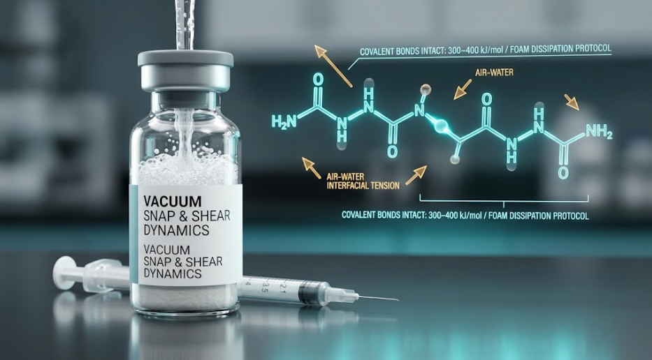
Managing an aggressive peptide reconstitution vacuum is a common challenge for laboratory researchers and bench technicians. You insert the needle expecting a gentle drip down the glass wall. Instead, negative pressure inside the vial yanks the plunger forward. The diluent violently jets into the dry powder, generating white foam and bubbles within seconds.
The immediate concern is catastrophic degradation. Online research forums often warn that peptide bonds snap under strong mechanical impact. However, biophysical literature paints a very different picture. Did that sudden vacuum snap ruin your sample, or is the risk exaggerated? Below, we examine the mechanics of the peptide reconstitution vacuum, shear stress limitations, and how to verify compound viability.
Quick Summary: Peptide Reconstitution Vacuum at a Glance
Quick Answer: A strong vacuum confirms a secure, hermetic seal from lyophilization. While rapid fluid entry creates foam and interfacial stress, it does not possess enough energy to break primary covalent peptide bonds. Letting the solution settle in refrigeration (2°C to 8°C) resolves micro-bubbles and preserves monomeric yield.
| Action Step | Correct Procedure | What NOT to Do |
|---|---|---|
| 1. Immediate Handling | Stop immediately. Remove the needle and set the vial upright on a flat surface. | Do not shake or roll the vial to dissolve the foam faster. |
| 2. Foam Dissipation | Place the vial in cold storage at 2°C to 8°C (36°F to 46°F) inside a secure Peptide Vial Case. Let it sit for 30 to 60 minutes. | Do not tap or flick the glass, as agitation creates secondary shear stress. |
| 3. Visual Clarity Check | Inspect the settled solution against light. The liquid should be crystal clear, water-thin, and free of visible particulate. | Do not use solutions that remain cloudy, opalescent, or show stubborn precipitation. |
| 4. Pressure Normalization | Insert a sterile EasyTouch 31G Syringe barrel without the plunger to equalize internal pressure before drawing a dose. | Do not leave unvented pressure imbalances that cause spraying on future needle entries. |
Why Are Peptide Vials Under Vacuum in the First Place?
The sudden suction is not a packaging defect. In fact, it is a deliberate outcome of pharmaceutical freeze-drying. A comprehensive overview from the National Center for Biotechnology Information (NCBI) on lyophilization principles details why pressure differentials exist:
- The Sublimation Cycle: Peptides are dissolved in formulation buffers, frozen solid, and placed under high vacuum. This low pressure enables sublimation, converting ice directly into water vapor.
- Stoppering Under Negative Pressure: When moisture levels drop below 3%, hydraulic shelves seal rubber stoppers into the vials under partial vacuum or an inert nitrogen backfill.
- Evidence of a Hermetic Seal: A strong peptide reconstitution vacuum confirms that the rubber septum and aluminum crimp remained airtight. In contrast, a vial lacking vacuum suction may have suffered seal failure, exposing the powder to atmospheric humidity.
The Physics of the "Snap": Mechanical Shear vs. Peptide Bonds
The primary concern during violent mixing is mechanical shear stress. When diluent jets into the glass vial, does it possess enough kinetic energy to break covalent bonds?
In short: Covalent peptide bonds do not shatter from liquid impact.
Peptide bonds linking amino acids require 300 to 400 kJ/mol of energy to break. Peer-reviewed investigations in biophysical protein stability literature confirm that fluid shear from syringe needles is far too weak to cleave covalent backbones. Even high-pressure homogenizers operating at thousands of psi rarely break primary amino acid sequences.
However, secondary and tertiary folding can still face physical stress:
- Short-Chain Peptides (<30 Amino Acids): Linear molecules like BPC-157, TB-500, and GHK-Cu lack delicate tertiary folds. Their biological activity depends on simple primary sequences. As a result, they are structurally resilient against rapid fluid impact.
- Long-Chain Proteins (>30 Amino Acids): Complex structures like human growth hormone (191 amino acids) rely on fragile hydrogen bonding and disulfide bridges. Intense turbulence can cause them to unfold partially, exposing hydrophobic residues.
What Does the Foam Actually Mean? (Interfacial Stress)
If chemical bonds stay intact, why do handling guidelines caution against rapid fluid flow? The true concern is the air-water interface.
When diluent shoots violently into a vial, it traps air bubbles. Peptides act as natural surfactants, aligning their hydrophobic segments toward trapped air bubbles. As noted in research on interfacial protein aggregation, this interaction causes several specific phenomena:
- Hydrophobic Exposure: Surface tension across micro-bubbles pulls folded domains open, triggering partial denaturation.
- Non-Covalent Aggregation: Exposed hydrophobic regions adhere to each other. This creates sub-visible dimers and trimers that lower the concentration of free, active monomer.
- Oxidative Exposure: Foaming significantly increases air exposure. If residues like methionine or cysteine are present, rapid aeration accelerates oxidative degradation over subsequent storage weeks.
Thus, while an abrupt peptide reconstitution vacuum pull will not sever covalent bonds, excessive foam reduces monomeric yield if handled carelessly.
What About "Adding Too Much Water"?
Another frequent bench error is injecting excess diluent—such as introducing 3 mL of Bacteriostatic Water instead of 1 mL or 2 mL.
Over-dilution never destroys peptide mass. A 5 mg vial contains 5,000 mcg of active material regardless of whether it is mixed with 1 mL or 4 mL of liquid. Only the concentration per unit volume shifts:
- Adjusted Volume: A diluted concentration simply requires drawing a larger volume on your syringe barrel to deliver the target microgram dose.
- Preservative Stability: Bacteriostatic water contains 0.9% benzyl alcohol. Adding extra diluent maintains this exact 0.9% concentration, ensuring reliable antimicrobial protection over its normal 28-day window.
- Volumetric Considerations: Larger injection volumes may require higher-capacity syringe barrels and can increase localized subcutaneous tissue distension.
How to Prevent Vacuum-Snap on Future Reconstitutions
To control the peptide reconstitution vacuum and prevent turbulence completely, follow these reliable techniques:
- Equalize the Diluent Vial: Inject an equivalent volume of room air into your bacteriostatic water vial before withdrawing liquid. This prevents hydraulic resistance.
- Control the Plunger: Keep your thumb braced firmly against the syringe plunger as you pierce the peptide vial. Never release your grip. Counter the internal vacuum pull so diluent enters at a steady drip.
- Aim for the Glass Wall: Angle the needle at 45 degrees toward the inner glass wall. Allow the fluid to trickle gently down the sidewalls into the bottom of the vial.
- Allow Passive Solvation: Never spray diluent directly into the lyophilized cake. Allow rising liquid to hydrate the powder gently from beneath, then swirl the vial in slow circles until clear.
If an unexpected peptide reconstitution vacuum snapped your syringe plunger, your vial is almost certainly safe. Let the foam settle quietly in refrigeration, verify clarity, and control plunger resistance carefully during your next preparation.
Understanding how a peptide reconstitution vacuum operates ensures you never mistake normal lyophilized negative pressure for sample destruction. By controlling liquid transfer speeds, avoiding violent agitation, and verifying clarity, you protect compound integrity. For further guidance on aseptic techniques, review our complete How to Reconstitute Peptides Guide and calculate required draw volumes with our Peptide Calculator.
← View all peptide guides, research & protocols in our Blog Archive